

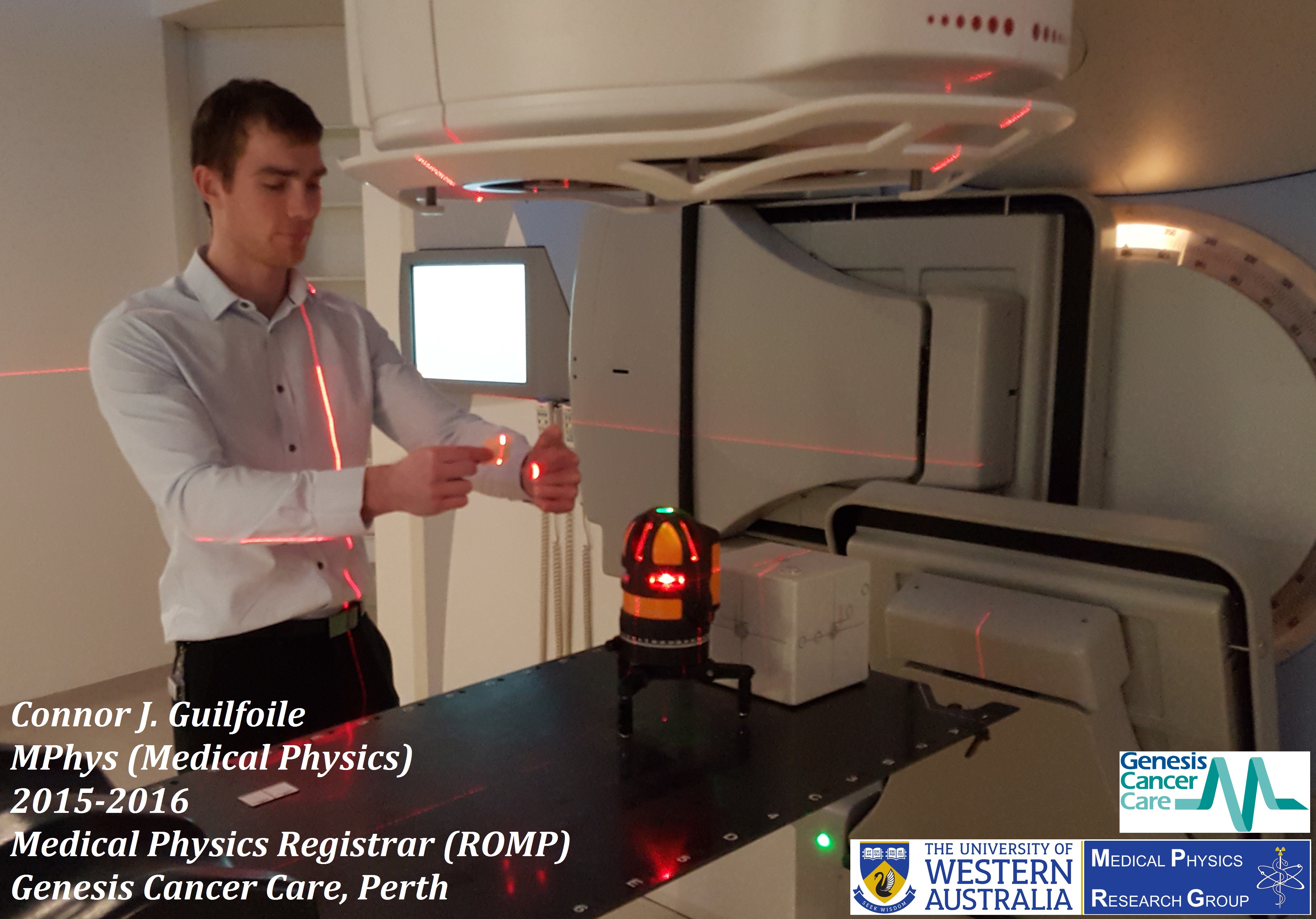
We are pleased to congratulate Mr. Connor James Guilfoile for being awarded a Master of Physics –Medical Physics from the School of Physics, University of Western Australia. Connor was one of our hardworking students and a few weeks after graduation, he was accepted to enter the Radiation Oncology TEAP program at Genesis CancerCare WA.
The title of his Masters project was: “Investigation of MAR (GE CT metal artefact reduction algorithm) and its suitability for use in radiotherapy treatment planning”.
His research was carried out under supervision of Mr. Peter Rampant and Assoc/Prof Michael House.
Part of Connor’s research outcome is published in the Australasian Physical & Engineering Sciences in Medicine (2017) 40:385–394. (Link to his paper)
The manuscript title is: “The impact of smart metal artefact reduction algorithm for use in radiotherapy treatment planning”
Connor kindly accepted to answer a few questions about his experience in our Medical Physics research Group.
 Click on the image to enlarge.
Introduction, your current position and role:
I studied for four years at Curtin University, where I completed my Bachelors and Honours in Physics and Mathematics.
Next, I went to the University of Western Australia to study my Masters of Physics (Medical Physics) for one and a half years.
Upon completion of my Masters degree, I successfully acquired a position at Genesis CancerCare WA. My current role is a Medical Physics Registrar. For the next three years I will be completing the Radiation Oncology Medical Physicist Training and Assessment Program (ROMP TEAP) offered by the ACPSEM (governing body for Medical Physicists in Australia). Once completed, I will become a qualified Radiation Oncology Medical Physicist.
What did you enjoy most about UWA?
The grounds and facilities were definitely a highlight, it is a great location with plenty of places to wind down and de-stress.
Can you give us your top three reasons to study Medical Physics?
- If you want to make a difference to the lives of people with cancer.
- If you enjoy a mental challenge.
- If have a passion for maths and science.
How do you feel you have made a difference in your field of research?
My Masters project investigated Metal Artefact Reduction (MAR) algorithms. These algorithms are designed to improve the image quality of CT images corrupted by metal artefacts. Using MAR enables a more accurate dose to the patient, leading to better patient outcomes. My project allowed me to investigate the pros and cons of using MAR algorithms and their usefulness in a clinical setting.
What is your best advice to current students and Medical Physics applicants?
Study hard and learn as much as you can. You’d be amazed at how useful prior knowledge can be, even when it seems completely irrelevant at the time.
Also get hands on experience; it is so beneficial to see protocols and procedures in action.
Lastly, make time for sport and recreation. Being able to stay stress-free is very important.
Once again we congratulate Connor for his great achievements and wish him all the best for his future career.
 Click on the image to enlarge.
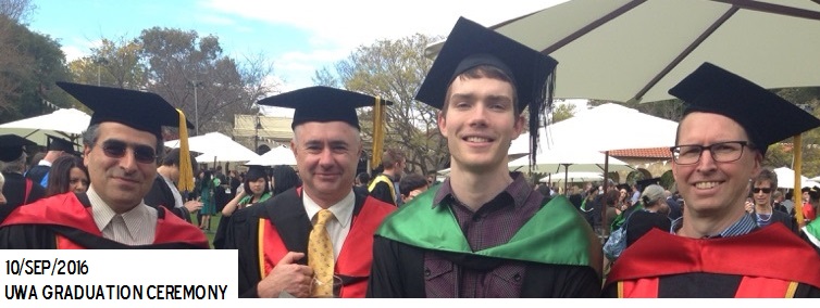
Connor’s graduation ceremony. from left to right: Assistant Professor Pejman Rowshanfarzad, Professor Martin Ebert, Mr. Connor Guilfoile, and Associate Professor Michael House
Here is the abstract of Connor’s thesis:
The presence of metal artefacts in Computed Tomography (CT) scans poses a number of issues for radiation oncology departments. It causes a loss of anatomical information and incorrect Hounsfield unit values add inaccuracies to dose calculations, providing suboptimal treatment for patients. Recently, the metal artefact reduction (MAR) algorithms have been developed to combat these problems.
One of these is known as: “Smart Metal Artifact Reduction (MAR)” released commercially by the General Electric Healthcare Company. The aim of this study was to provide a qualitative and quantitative analysis of the GE-MAR and determine its usefulness in a clinical setting. This was achieved by performing a number of studies using both patient and phantom data, noting any improvements in Hounsfield unit values and dosimetry with the GE-MAR enabled.
Findings from this study indicate qualitative improvements in severity of the streak artefacts produced by metals, allowing for easier patient contouring. Additionally, the GE-MAR managed to recover anatomical information which was previously lost due to artefact. Further findings in phantom data conveyed an improvement in Hounsfield unit value with GE-MAR corrected data, leading to more accurate treatment planning point dose calculations.
For future research, it is recommended that the GE-MAR is tested for a variety of patient implants not considered in this study including: plates, pacemakers and dental fillings. Further, gold standard dose distributions of patient treatment plans would be ideal and allows one to determine if in fact the GE-MAR images provide a more accurate dose distribution. Summarising this study, the GE-MAR was found to be a useful tool and should be implemented in clinical environments.
 Click on the image to enlarge.
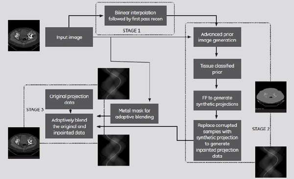
A flow chart of the GE Smart Artifact Reduction (MAR) algorithm