

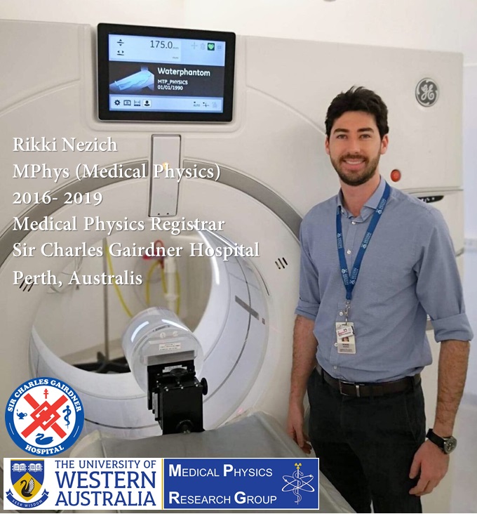
In this post, we are going to introduce one of our brilliant Medical Physics graduates, Mr. Rikki Nezich.
Rik started his MPhys (Medical Physics) at UWA in 2016. He had a First Class Honours Degree in Biophysics before starting his masters course of study.
Rik’s research project was on “Quantitative imaging of gamma-ray emitting radionuclides on a preclinical positron emission tomography (PET) scanner” supervised by Adj. Prof. Roger Price, and Mr. Steven Crossley from Sir Charles Gairdner Hospital (SCGH), WA, Perth.
Rik was working at Medical Technology and Physics Department at SCGH for a few years before starting his masters and is now a Diagnostic Imaging Medical Physics Registrar at SCGH.
Here are comments from his principal supervisor, Adjunct Professor. Roger Price:
“Rik joined Medical Technology & Physics, SCGH as both a staff member and a student at a time when preclinical radiopharmaceutical imaging systems were being installed in WA at the Perkins Institute. Students in MTP (and indeed in most Hospital departments) are obliged to be ‘self-starters’ and to interact with expert but very busy professionals. Rik chose a preclinical-PET topic for his (First Class) Honours Degree in Biophysics and virtually single-handedly mastered the math and computations required to examine the effects of positron drift and other phenomena on image quality – using a range of positron-emitting isotopes. He extended this work in his choice of project for a Masters of Medical Physics. Unlike the familiar 18F and 11C, some positron-emitting radioisotopes with emerging applications for targeted PET molecular imaging – particularly suited to theranostic applications – also co-emit prompt gamma rays. The effect of these singleton quanta on 511-coincidence detection and consequently image quality has been relatively little studied. Rik has made an important contribution to this field (again, virtually single-handedly) and hopefully will carve out time to extend the work, which will become increasingly relevant as preclinical (and eventually clinical) PET imaging reaches towards theoretical limits for temporal and spatial resolutions.”
Rik kindly accepted to answer a few questions about his experience in our Medical Physics research Group.
Introduction and your current position and role:
Hi, my name is Rik; I’m a Diagnostic Imaging Medical Physicist currently working in the Department of Medical Technology and Physics at Sir Charles Gairdner Hospital. My role is to provide diagnostic medical physics and radiation safety services across four hospitals in Perth. I’m also training as a registrar in the ACPSEM TEAP program in order to become an accredited Medical Physicist. I completed my Master of Medical Physics degree at the University of Western Australia in mid-2019, which I studied part-time while working in my current job.
What did you enjoy most about UWA, and Medical Physics research group?
Probably the most enjoyable aspect of studying at UWA was having the opportunity to meet so many excellent and inspirational people, in the Medical Physics research group, and through various UWA student associations and events. I was also grateful to benefit from an exceptional level of support and mentoring from the staff and students in the research group.
Can you give us your top three reasons to study Medical Physics?
1- As a trained Medical Physicist, your specialised knowledge and skills are crucial to the delivery of safe and high quality healthcare.
2- Medical Physicists are closely involved in the clinical implementation of exciting new medical technologies.
3- Medical Physicists are generally well-compensated for their skills and efforts, and have meaningful careers which benefit patients and society.
How do you feel you have made a difference in your field of research?
My research project was to investigate the impact of prompt gamma-ray emissions on the quantitative accuracy of positron emission tomography (PET) imaging. My research showed that the accuracy and precision of PET images are impaired when utilising certain gamma-ray emitting radionuclides, and demonstrated how these effects can be partly mitigated with the selection of appropriate image acquisition and reconstruction parameters. My research also highlights the need for data correction strategies to be further developed and implemented on commercial PET scanners.
What is your best advice to current students and Medical Physics applicants?
Do not underestimate the importance of having good communication skills –Medical Physicists are required to cooperate with many different people across multiple working groups and specialties. Also, take advantage of networking opportunities to meet Medical Physicists working in academia and industry. In my experience, this can be incredibly useful for getting a great job in Medical Physics.
Abstract of Rikki’s thesis:
Introduction: Positron emission tomography (PET) is a molecular imaging technique by which a radioactive tracer molecule is administered into a living organism to measure the state of metabolic processes in the body. In medicine, quantitative PET imaging is central to the clinical diagnosis and management of cancer; as well as to the preclinical research and development of novel pharmaceutical drugs, internal radionuclide therapies, and imaging tracers. A number of unconventional (non fluorine-18 or carbon-11) positron emitting radionuclides are increasingly being utilised for applications that include immuno-PET and ‘theranostics’ imaging. However, many of these non-traditional radionuclides exhibit complex radioactive decay schema involving the emission of gamma rays, which are known to potentially impair the quantitative accuracy of PET imaging. The aim of this project was to investigate the quantitative accuracy and precision of PET imaging with a preclinical small-animal PET scanner (SuperArgus, Sedecal), for several gamma-ray emitting radionuclides of biomedical importance (copper-61, gallium-68, bromine-76, rubidium-82, yttrium-86, and iodine-124), in comparison to fluorine-18.
Materials and methods: The quantitative accuracy and precision of PET data acquired with the aforementioned gamma-ray emitting radionuclides was measured using a computational Monte Carlo simulation model of a SuperArgus PET scanner. The scanner model was purpose-built for this investigation using the Monte Carlo simulation platform ‘Geant4 Application for Tomography Emission’ (GATE), and was validated against a physical version of the scanner in terms of system sensitivity, count rate behaviour and spillover ratios. Using the simulation model, simulated PET images of a NEMA NU4 image quality phantom were obtained for each of the radionuclides of interest, to evaluate the following metrics of quantitative image accuracy and precision: Spillover ratios in water (SORwater) and background air (SORbkg), contrast ratios (CR), signal-to-noise ratios (SNR), and mean-signal ratios relative to fluorine-18 (MSRF-18).
Results: Absolute sensitivity, count rates and spillover ratios of the simulated scanner were validated against those of the physical scanner. In comparison to fluorine-18, spillover ratios in water and background air were most impacted with bromine-76, yttrium-86, and iodine-124. Contrast ratios were significantly degraded with yttrium-86. Signal-to-noise ratios were reduced for all radionuclides in comparison to fluorine-18. Quantitative accuracy was generally improved with use of the narrow energy window (400-700 keV), 2D filtered backprojection reconstruction, and with applied corrections for randoms and scatter.
Conclusion: PET quantitative accuracy is impacted by gamma-ray emission with the radionuclides bromine-76, yttrium-86, and iodine-124; in comparison to fluorine-18. Radionuclide-specific activity calibration factors should be applied for PET imaging with these radionuclides.
Rik’s research poster describing his Master’s work won an award for ‘Best Poster Presentation – Imaging Category‘ in EPSM conference-2019, Perth.
Rik has created a website and has shared his software for CT image QA named “Precision Radiation Services“.
We wish Rik all the best in his future career as a Medical Physicist.
![]()